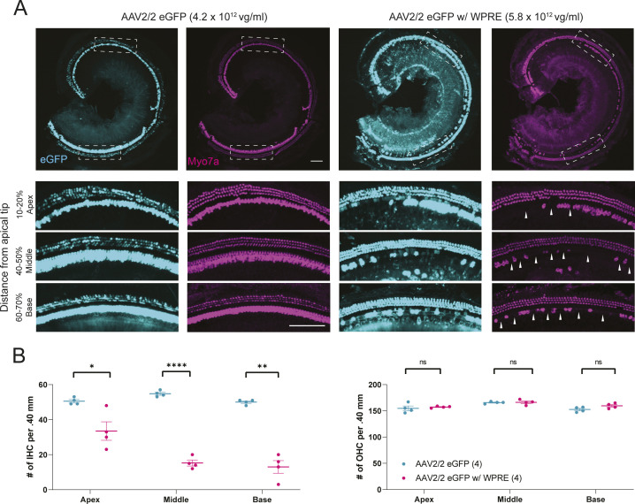
Figure S3. Immunohistochemistry at 10 wk of age after RWM + CF inoculation with eGFP marker vectors.
(A) Whole-mount immunostaining of WT controls that received AAV2/2 eGFP and AAV2/2 eGFP w/WPRE. Whole-mount dissection and immunostaining for Myo7a (magenta) and imaging for native eGFP (cyan) were performed at 10 wk of age. Scale bars represent 100 μm, and white dashed boxes represent regions of high-magnification image capture for cell counting. Arrowheads: hair cell loss. (B) Cell counting of WT controls that received marker vectors. AAV2/2.miSafe.eGFP-injected mice showed no evidence of hair cell loss. AAV2/2.miSafe.eGFP.WPRE-injected mice showed hair cell loss at 10 wk of age, consistent with the observed increase in auditory thresholds (Fig 2C). Hair cell loss was predominantly observed in inner hair cells, whereas outer hair cells were intact. Data are means ± SEM. N mice per experimental condition are indicated in parentheses. Statistical analysis was performed by Welch’s t test. Inner hair cell comparisons: apex, P = 0.046; middle, P < 0.0001; and base, P = 0.002. ****P < 0.0001; **P < 0.01; and *P < 0.05.

