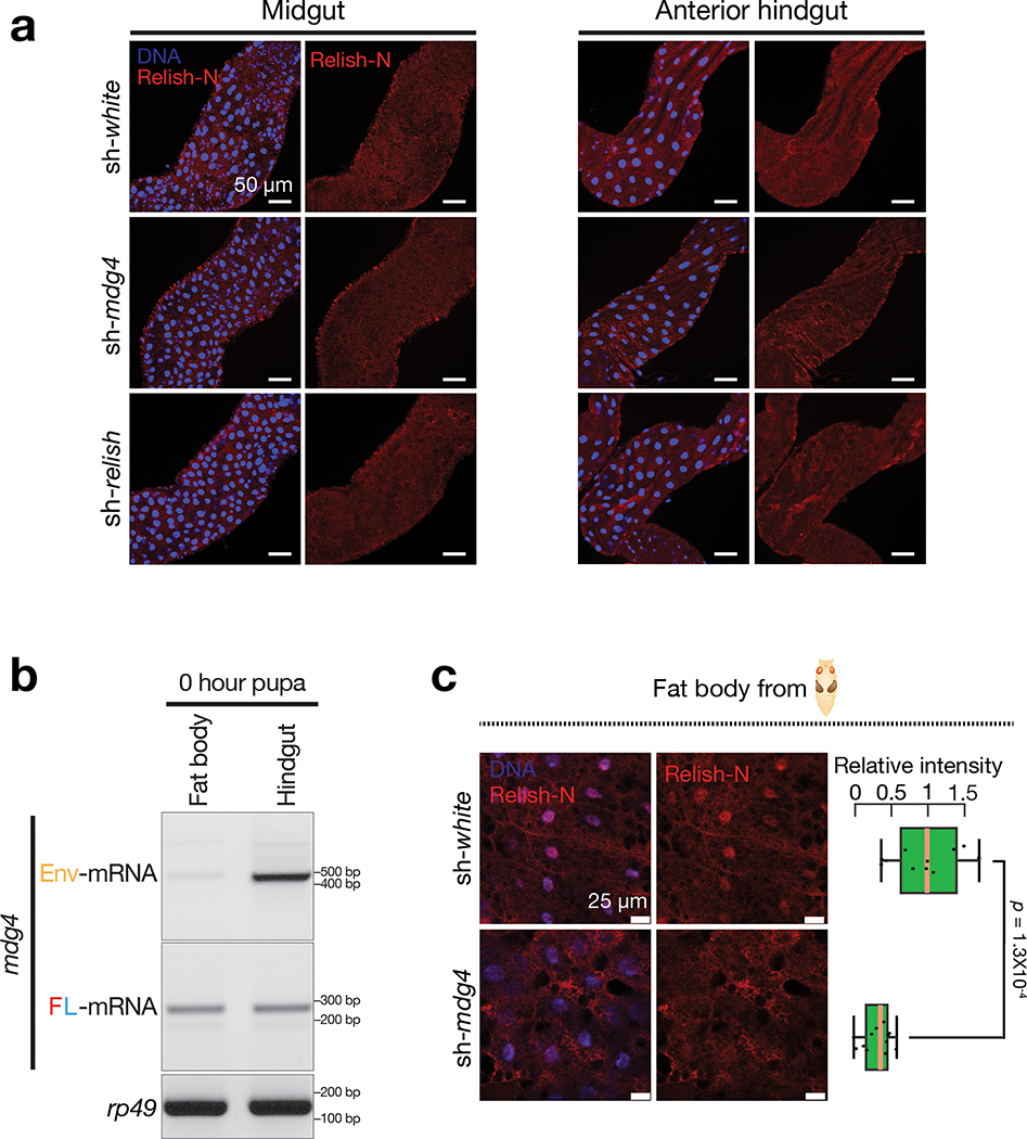
Extended Data Fig. 9 |. mdg4 products promote the translocation of Relish-N into nucleus.
a, By performing immuno-staining with the Relish-N antibody, which can detect both full-length and N-terminal fragment of Relish, very low–if any–signals were detected in midgut and anterior part of hindgut from sh-white, sh-mdg4, or sh-relish early pupae. b, RT-PCR to examine the levels of mdg4 full-length and Env transcripts in fat body cells. Two independent biological replicates were performed for a and b. c, Immuno-staining to detect the nuclear Relish-N signals in the fat body cells from sh-white and sh-mdg4 early pupae. While the animals for Fig. 7 were raised in germ-free condition, the pupae examined for this figure were non-germ-free. In both conditions, silencing mdg4 resulted in a significant decrease of the nuclear Relish-N in fat body cells. Box plots report the minimum, maximum, median, and interquartile ranges of the data. A two-tailed t-test was used to compare the relative intensities of each genotype. The data were collected from 3 individual animals per genotype.

