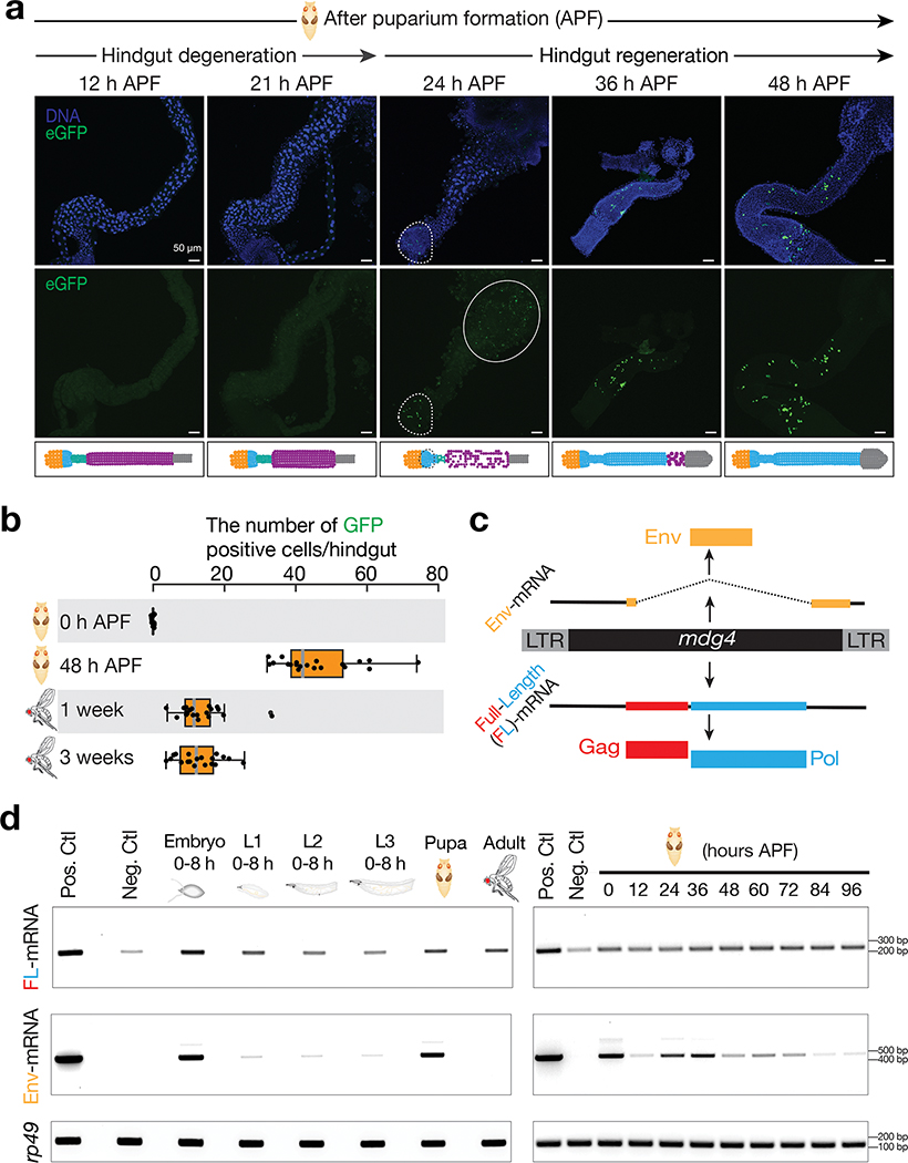
Fig. 2 |. mdg4 selectively mobilizes in the regenerating tissues during metamorphosis.
a, mdg4 mobilization in Drosophila during hindgut development at the pupal stage via eGFP transposition reporter. No eGFP is expressed during hindgut degeneration (12 h and 21 h APF). eGFP positive cells can be detected in the newly formed hindgut (24 h, 36 h, and 48 h APF). Dashed circle highlights eGFP signals. Solid circle depicts autofluorescence from the dying cells. Extended Data Figure 2a depicts the cell-type dynamics of hindgut during metamorphosis. b, The box plot shows the number of eGFP-positive cells per fly from mdg4 transposition reporter at different stages (N = 20). Box plots report the minimum, maximum, median, and interquartile ranges of the data. c, Diagram depicts the transcripts and proteins from mdg4. d, RT-PCR experiments to measure the expression of full-length and Env mRNAs from reporter-carrying flies. Pos. Ctl.: positive control, ovaries with Piwi being depleted in follicle cells. Neg. Ctl.: negative control, ovaries with mdg4 being depleted in both germ cells and follicle cells. APF: after puparium formation. Similar findings were made when using RNA-seq to quantify the full-length and Env mRNAs from endogenous mdg4 (Extended Data Fig. 4). Three independent biological replicates were performed.

