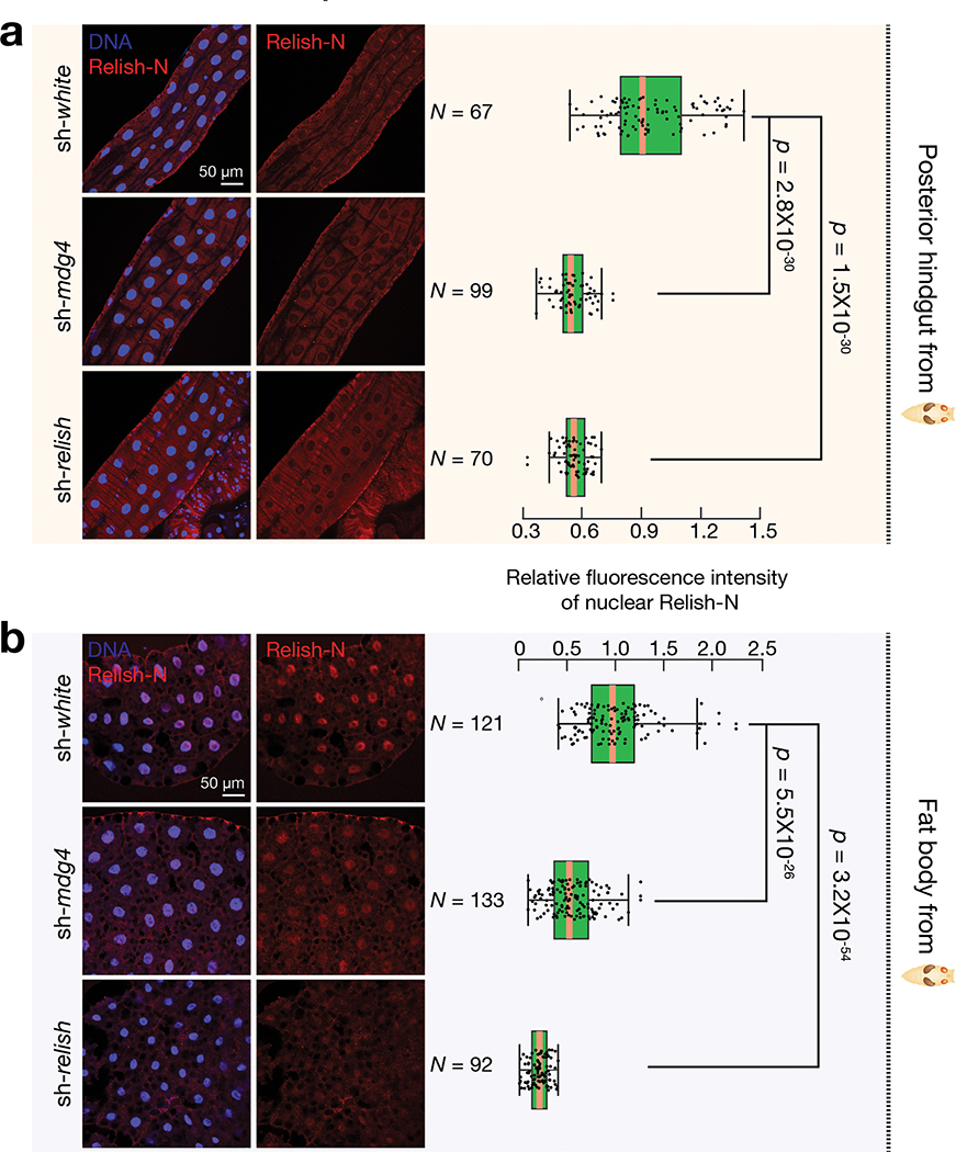
Fig. 7 |. mdg4 triggers Relish activation in both hindgut and fat body at pupal stage.
a, Immunostaining to detect the nuclear Relish-N signals in the posterior part of hindgut from sh-white, sh-mdg4, or sh-relish early pupae. The box plot shows the relative fluorescence intensity of nuclear Relish-N signals in the posterior part of hindgut. b, Immunostaining to detect the nuclear Relish-N signals in the fat body cells from sh-white, sh-mdg4, or sh-relish early pupae. Flies were raised on germ-free condition; Similar findings were made on non-germ-free condition (Extended Data Fig. 9c). The data were collected from 2 biological replicates with 3 animals per replicate. The box plot shows the relative fluorescence intensity of nuclear Relish-N signals in the fat body cells. Silencing mdg4 resulted in a significant decrease of the nuclear Relish-N in both hindgut and fat body cells. Box plots report the minimum, maximum, median, and interquartile ranges of the data. Two-tailed t-tests were used to evaluate the difference between each pair of groups.

