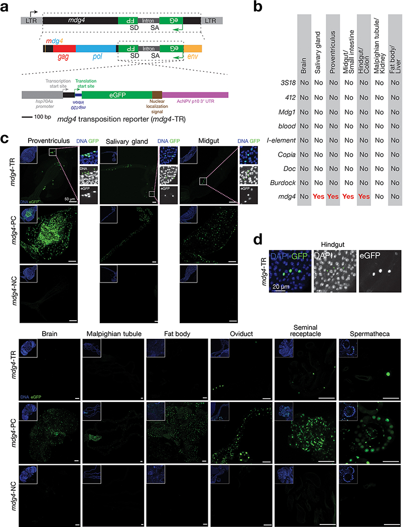
Extended Data Fig. 1 |. Monitoring retrotransposon mobilization in somatic cells via a transposition reporter.
a, Detailed schematic design of eGFP transposition reporter to monitor mdg4 mobilization. b, Summary of mobilization events from different somatic tissues for 9 retrotransposon families, as assayed by corresponding eGFP reporter. No: no eGFP positive cells are detected; Yes: eGFP positive cells can be detected. c, Detecting eGFP signals in somatic tissues from positive control, negative control, and mdg4 transposition reporter in 2–4-day-old adult flies. Note: Positive control construct gives low number of eGFP positive cells in brain and malpighian tubules, indicating that transcription of mdg4 is suppressed in these tissues. Three independent biological replicates were performed. d, Zoom-in display of the box region in Fig. 1b. In DAPI channels, green arrows point to the nuclei that have GFP expression.

