
Fig. 4. PEX13 adopts two transmembrane orientations.
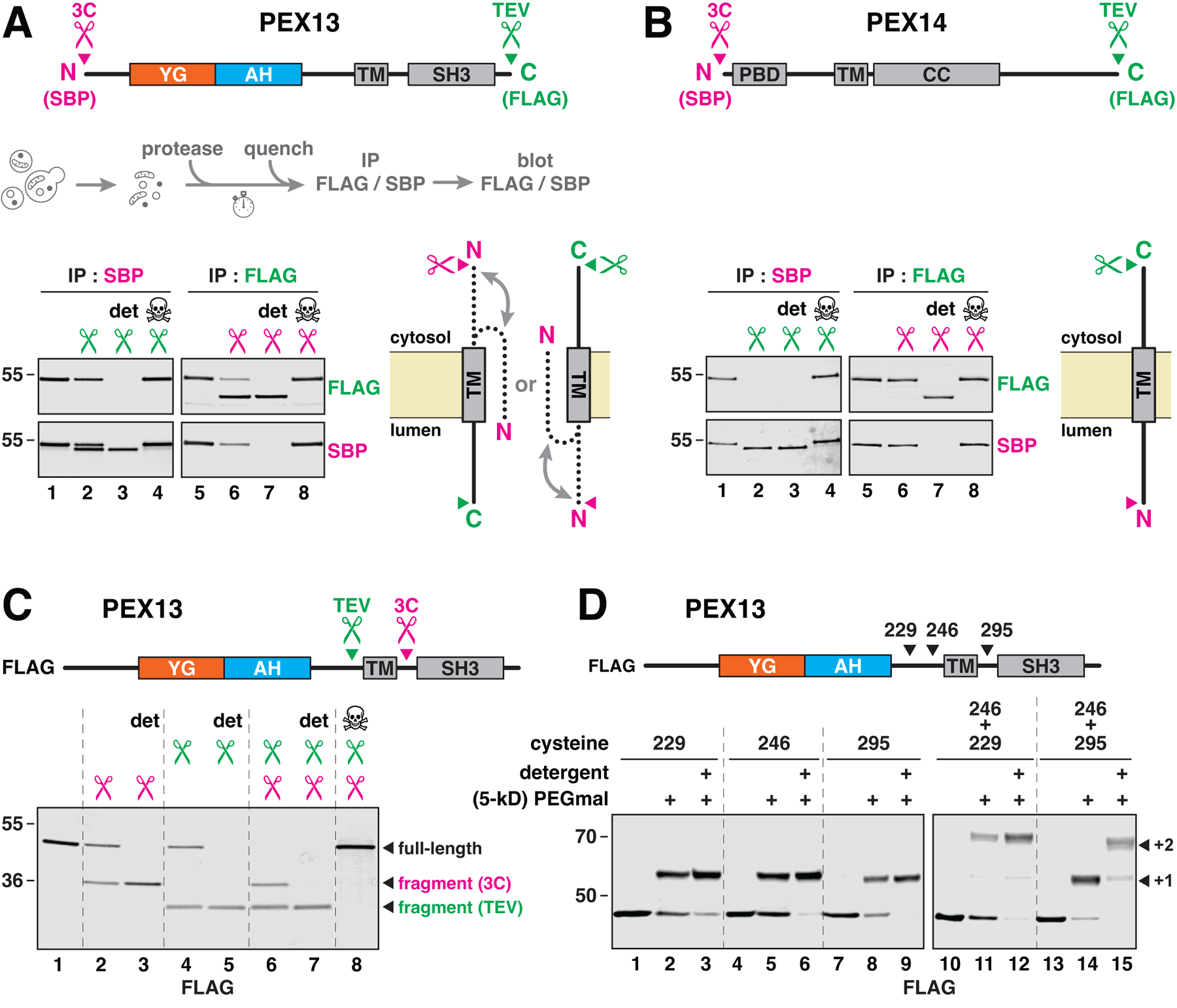
(A) Membrane topology determined by protease protection. The indicated protease-cleavage sites (scissors) and epitope tags were introduced into yeast PEX13. Membranes containing this protein were treated with protease with or without detergent (det), the reactions were quenched with NEM, and PEX13 was immunoprecipitated (IP); cleavage was visualized by immunoblotting (see scheme). Where indicated (skull and crossbones), NEM was added before the proteases. The position of the C terminus is deduced from cleavage with TEV protease and immunoprecipitation by the N-terminal SBP tag, and the position of the N terminus is deduced from cleavage with 3C protease and immunoprecipitation by the C-terminal FLAG tag. The two inferred orientations of PEX13 are depicted on the right; the N terminus can face either side, whereas the C terminus is fixed in one of two orientations. (B) Same as in (A), but for PEX14. PBD, PEX5- binding domain; TM, transmembrane segment; CC, coiled-coil oligomerization domain. (C) Same as in (A), but with protease sites flanking the TM and a FLAG tag as shown. (D) TM orientation determined by modification of flanking cysteines with membrane-impermeable PEGmal. One or two cysteines were introduced into FLAG-tagged PEX13, as indicated. Membranes were treated with 5-kD PEGmal and then quenched with excess cysteine; single (+1) or double (+2) modification was visualized by immunoblotting.