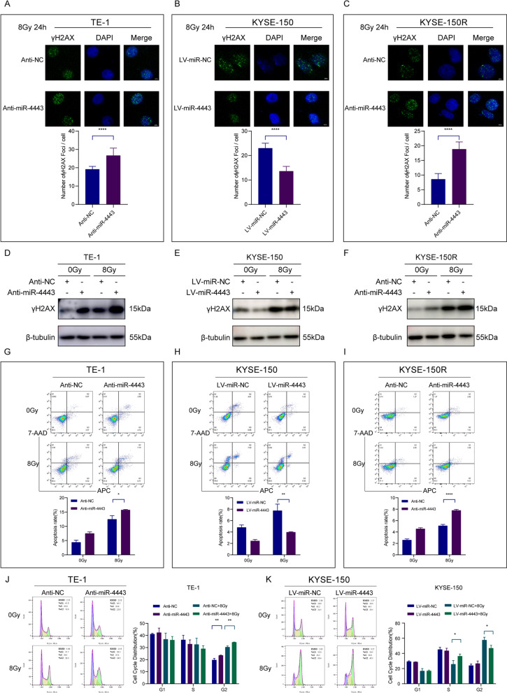
Fig. 3.
Knockdown of miR-4443 enhanced the radiosensitivity of ESCC cells. A–C Representative immunofluorescence images of nuclear γ-H2AX foci (cell nuclei: blue; γ-H2AX foci: green) in the indicated cells after 24 h of 8 Gy radiation. Scale bar: 4 µm. D–F Western blot analysis of γ-H2AX protein expression levels in the indicated cells after 24 h of 8 Gy radiation. G–I Evaluation of the apoptosis rates of ESCC cells with altered expression levels of miR-4443. J, K Cell cycle distribution in the indicated cells after 6 h of 8 Gy radiation. Data are presented as the mean ± SD; n = 3 independent experiments. (*p < 0.05, **p < 0.01, ***p < 0.001, ****p < 0.0001)

