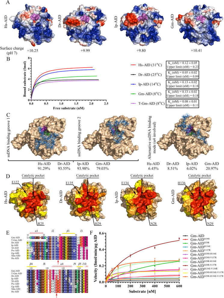
Fig. 5.
Structural modeling and mutagenesis to determine the basis of Atlantic cod AID lethargy. A Predicted surface topology of Gm-AID was compared to that of other AID orthologs. Positive, neutral, and negative residues are colored blue, white, and red, respectively. The putative catalytic pocket is colored in purple. Surface charge (at pH 7.00) is shown below each model. B The electrophoretic mobility shift assay (EMSA) was conducted to compare global ssDNA binding affinity of AID orthologs. Estimated Kd and upper limits show no significant difference among AID orthologs. C Docking of ssDNA on the surface of the Gm-AID model revealed the presence of the two main ssDNA binding groove 1 and 2 previously identified in Hs-AID, as well as alternative ssDNA binding mode which involved the α4 region. The contribution of different binding modes is shown for each AID ortholog. D Interactions between AID residues and ssDNA are shown as heatmaps. Amino acid residues interacting with substrate in 50–100%, 30–50%, 15–30%, 5–15%, 0–5%, and 0% of docking events are shown in red, dark orange, light orange, yellow, sand, and wheat colors, respectively. Shown with arrows are two potential amino acids that contribute to the increasing involvement of Gm-AID α4 and their counterparts in other AID orthologs. E Partial alignment of the AID orthologs surrounding Gm-AIDN29, Gm-AIDH136, and Gm-AID.V137 residues. The approximate secondary structure of α-helical (α), β-strand (β), and loop regions (l) are shown on top of the alignment. F The catalytic rate of Gm-AID mutants was compared to that of wildtype Gm-AID through Michaelis–Menten kinetics. Data are represented as mean ± SEM (n = 4)

