
Fig. 2. Macular Bruch’s membrane elastin layer degradation in AMD.
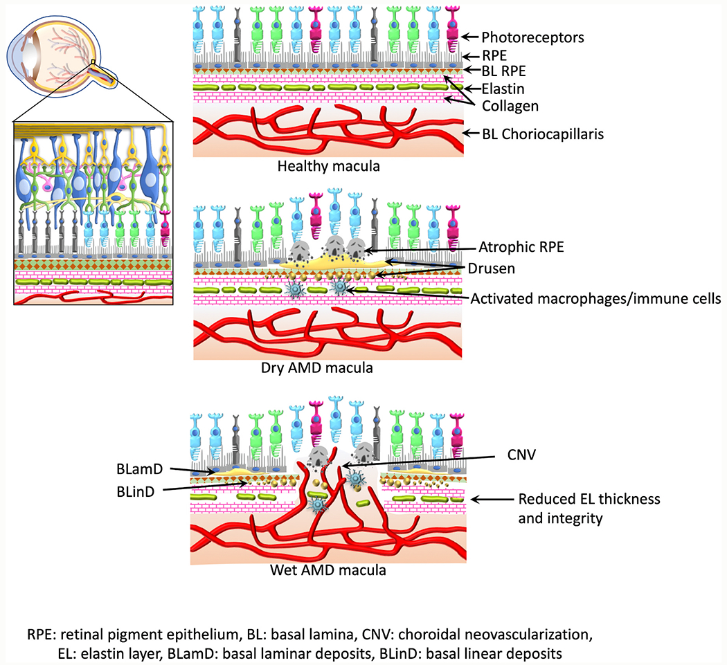
Figure indicates schematic representation of eyeball, retina, and macula during health and AMD. Reduced macular elastin layer integrity and thickness have been reported in AMD patients compared to the age matched healthy subjects; BrM elastin layer thinning, calcification, and porosity were more evident and were corresponding to the distribution of CNV lesions in wet AMD macula.