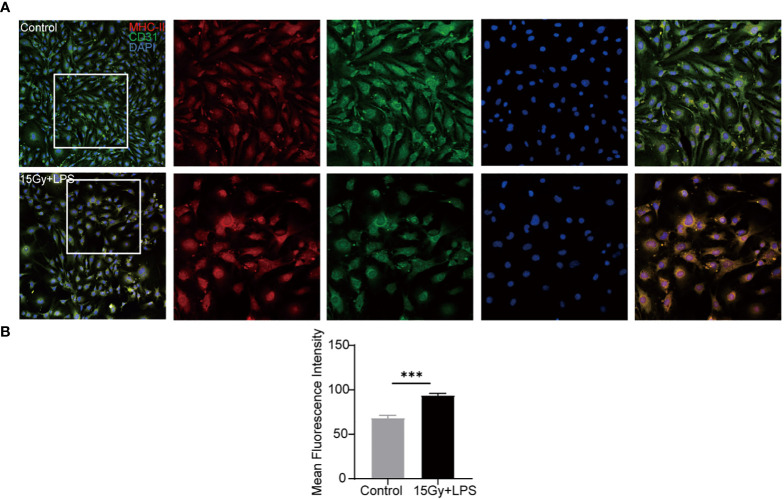
Figure 6.
Changes in the expression of MHC-II molecule after endothelial cell injury. Endothelial cells were irradiated (15Gy) and stimulated with LPS (100 ng/ml) for 24 hours followed by immunofluorescence staining with anti- mouse MHC-II molecule and anti- mouse CD31 antibodies. Red: MHC-II molecule, green: CD31, blue: nucleus. (A) Image J software analyzed the mean fluorescence intensity of MHC-II molecule in cells (n=4, *** P<0.001) (B).

