Figure 3.

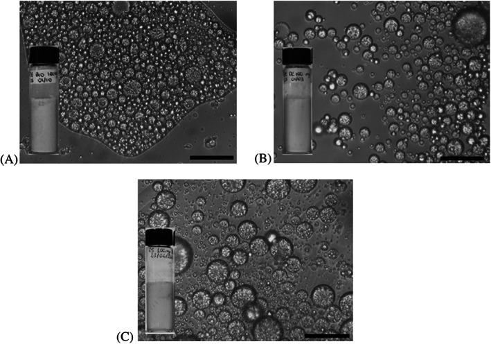
Optical microscopy images of fresh samples (scale bar is 25 μm) and corresponding visual appearance of (A) 05PE_GDPBIO, (B) 05PE_GDPC1, and (C) 05PE_GDPC2.

Optical microscopy images of fresh samples (scale bar is 25 μm) and corresponding visual appearance of (A) 05PE_GDPBIO, (B) 05PE_GDPC1, and (C) 05PE_GDPC2.