
FIGURE 9.
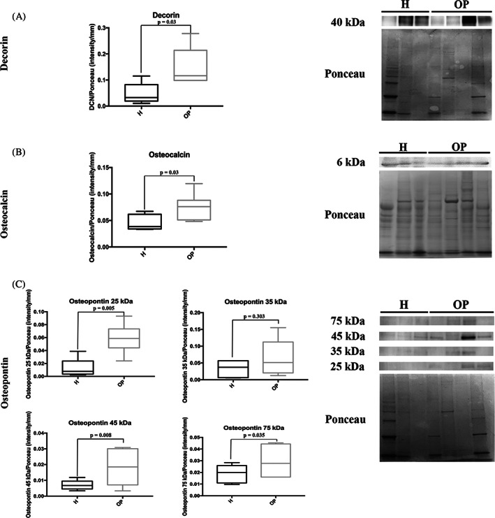
Representative Western blotting image and densitometry for (A) Decorin, (B) Osteocalcin, and (C) Osteopontin in healthy (H) and osteoporotic (OP) tissues (the same Ponceau staining was used to normalize proteins analyzed at different molecular weight on the same membrane)