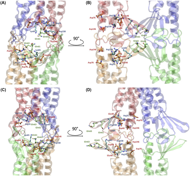
FIGURE 3.

Representations of relevant residues in the two pore models. (A,B) Apical/basolateral A and lateral, B, views of the Pore I configuration. The β‐barrel arranged by the ECLs of the four protomers is visible. (C,D) Apical/basolateral, C, and lateral, D, views of the Pore II configuration. The reverse orientation of the pore‐lining residue compared to the Pore I is visible. The pore‐lining residues are indicated for two opposing protomers with respect to the paracellular plane. Acidic residues are depicted in orange, basic residues in light blue, and neutral residues in green. Oxygen and nitrogen atoms are shown in red and blue, respectively.
