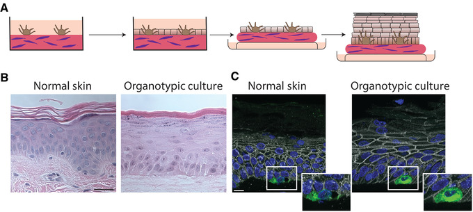
Figure 1.

Organotypic skin cultures model normal skin. (A) Schematic representing the general formation of 3D organotypic skin cultures incorporating melanocytes. A collagen‐fibroblast plug is created on which melanocytes are seeded and allowed to adhere for 24 hr. NHEKs are seeded onto the plug and allowed to adhere and reach confluence for 48 hr. The culture is then lifted to an air liquid interface inducing stratification and differentiation. (B) H and E stained FFPE samples from normal skin compared to an organotypic skin culture with incorporated melanocytes after lifting to an air liquid interface for 6 days. Organotypic skin cultures retain the layers normally present in skin, including a cornified layer and the presence of keratohyalin granules in the granular layer. Scale bar = 50 μm. (C) FFPE tissue sections from normal skin or organotypic skin cultures stained for Melan A to label melanocytes, plakoglobin to label cell borders, and DAPI. Melanocytes in the organotypic skin cultures are localized to the basal layer like normal skin. Scale bar = 20 μm. Abbreviations: NHEKs, neonatal human epidermal keratinocytes; H and E, hematoxylin and eosin; FFPE, formalin fixed paraffin embedded.
