
Figure 6.
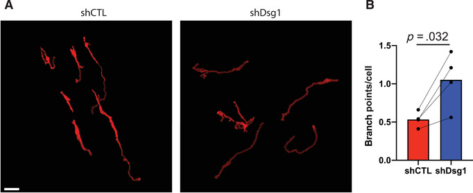
Measuring changes in melanocyte dendricity in organotypic skin cultures. Activation of the tanning response in melanocytes results in both an increase in pigment production, as well as an increase in melanocyte dendricity to promote transfer of pigment to the surrounding keratinocytes. Activation of the tanning response can be mediated by loss of keratinocyte desmoglein 1 (Arnette et al., 2020). To measure melanocyte morphology the melanocytes were transduced with a construct expressing tdTomato allowing visualization of the cells in the organotypic culture. To whole mount stain the organotypic cultures the epidermal equivalent was removed from the collagen plug and fixed in 4% paraformaldehyde and stained with DAPI before mounting to a slide. The entire thickness of the organotypic culture was then imaged using a Nikon A1R confocal microscope equipped with GaAsP detectors and 20× Plan‐Apochromat objective with a NA of 0.75. To measure changes in melanocyte morphology the SNT plugin for FIJI was used to generate 3D models of the melanocytes, and these were used to determine dendrite length and branches of the melanocyte dendrites. (A) Representative images of melanocytes in organotypic cultures with control (shCTL) or desmoglein 1 knockdown (shDsg1) NHEKs. Scale bar = 50 μm. (B) Quantification of the average number of branchpoints per melanocyte under each condition. NHEKs, neonatal human epidermal keratinocytes.