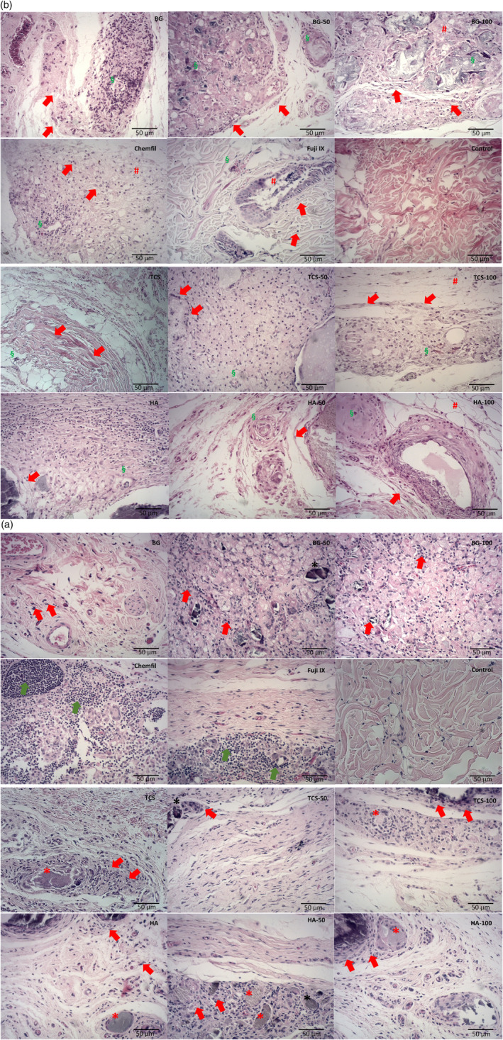
FIGURE 4.

(A) Hematoxylin & eosin‐stained representative light micrographs (40×), after 30 days, of the subcutaneous tissue adjacent to the material implant showing inflammatory cell infiltrate (red arrows) for all materials and its strontium replacements in different levels. In the Fuji and ChemFil samples an intense inflammatory cell concentration (green arrows) could be observed, and normal histology was observed in the negative control samples. Besides, detached material fragments (*) could be observed interposed within the connective tissue surrounded by a characteristic inflammatory chronic cell infiltrate representing a granulomatous foreign‐body reaction (n = 5 for each material at different time points). (B) Hematoxylin & eosin‐stained representative light micrographs (40×), after 180 days, of the subcutaneous tissue adjacent to the implant showing a mild inflammatory cell infiltrate with collagen fibers (arrows) indicating potential tissue repair. Tissue disorganization (#) was observed for all the 100% strontium‐doped groups, Fuji, and ChemFil. Moderate angiogenesis and mild fibrosis (§) were also evident in this period of analysis for the test groups, whereas a more normal histology was observed for the negative control. (n = 5 for each material at different time points)
