FIGURE 7.

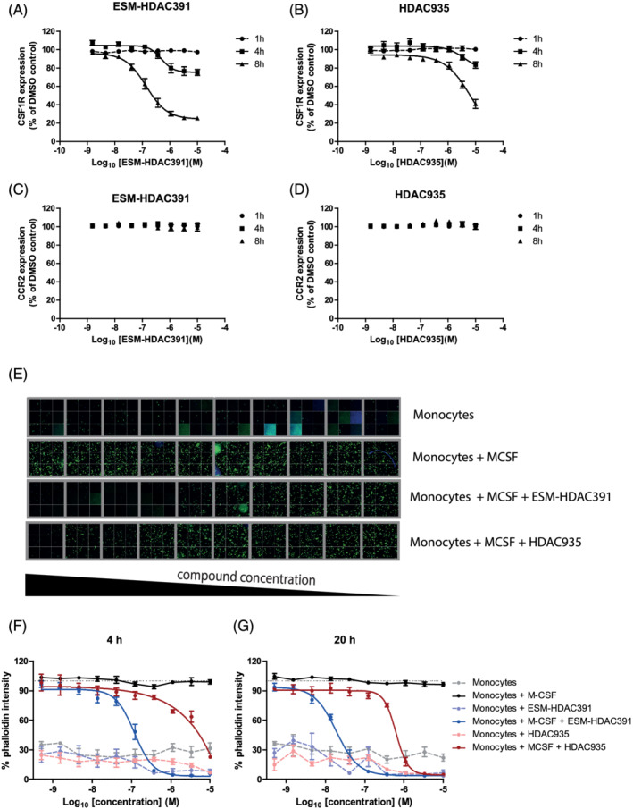
ESM‐HDAC391 downregulates CSF1R and affects monocyte differentiation. (A, B) Concentration‐ and time‐dependent reduction in CSF1R on monocytes incubated with ESM‐HDAC391 and HDAC935, respectively (n = 4). (C, D) Parallel expression of CCR2 levels on monocytes following incubation as above with ESM‐HDAC391 and HDAC935, respectively (n = 4). (E) Cytoskeletal F‐actin staining images following 48 hours differentiation of CD14+ monocytes in the presence of macrophage colony‐stimulating factor (M‐CSF) with an initial 4 hours exposure to ESM‐HDAC391 or HDAC935 (final concentrations titrated from 10 μM to 0.51 nM) followed by compound washout and continued incubation with M‐CSF in fresh media for the remaining 44 hours; representative data of 32 fields of views per well from 1 of the n = 4 replicate donors are shown. (F, G) Normalised quantification of cytoskeletal F‐actin staining following 48 hours differentiation of CD14+ monocytes in the presence of M‐CSF with an initial incubation with ESM‐HDAC391 or HDAC935 for 4 or 20 hours (respectively) followed by washout (n = 4)
