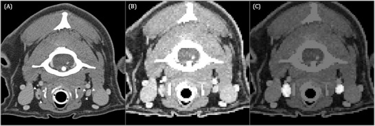
FIGURE 4.

Transverse contrast‐enhanced CT image acquired in sternal recumbency (window width = 450, level = 140, 120 kVp, 280 mA) (A) at the level of the caudal atlas of the same dog shown in Figure 3 depicting the medial retropharyngeal lymph nodes (white arrowheads). Input image for U‐Net (B) and corresponding output image (C). The pixels identified by U‐Net as medial retropharyngeal lymph nodes are highlighted in image (C)
