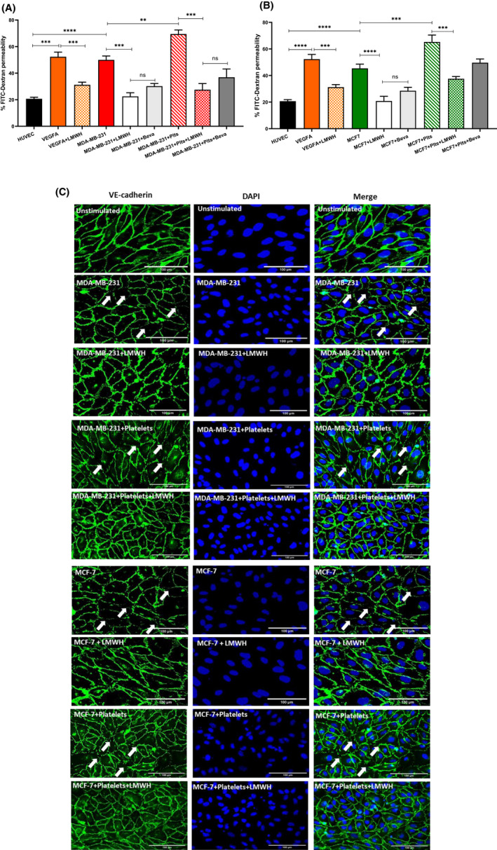
FIGURE 5.

Breast cancer cells induce enhanced EC permeability and transendothelial migration. Breast tumor supernatant from (A) MDA‐MB‐231 and (B) MCF‐7 cells either alone or co‐cultured with platelets was collected and applied to a confluent HUVEC monolayer on a transwell insert. EC permeability was assessed by leakage of 10 kDa FITC‐Dextran from the top chamber after stimulation. (C) Immunofluorescence staining for VE‐cadherin was performed on HUVEC after tumor stimulation with and without LMWH pretreatment. Arrows indicate areas with reduced HUVEC barrier integrity. Scale bar 100 μm. Data were analyzed with anova followed by Bonferroni t ‐test and p values < .05 were considered to be significant (*p < .05, **p < .005, ***p < .001).
