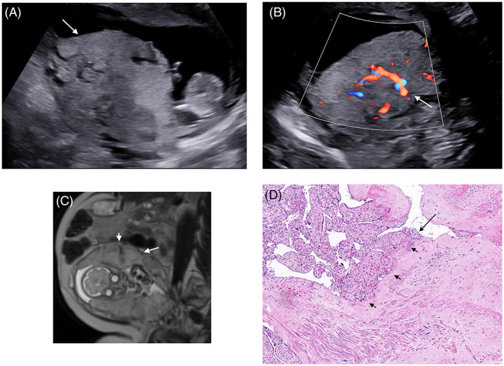
FIGURE 1.

Gray‐scale ultrasound at 12 weeks showing the posterior lobe of the placenta (arrow) with loss of the hypoechoic retroplacental (clear) zone, numerous large or irregular lacunae (A); color Doppler ultrasound shows a feeding vessel with flow velocity >20 cm/s (arrow) (B); MRI T2‐weighed image at 28 weeks showing the bilobed placenta with heterogeneous posterior lobe (arrow), multiple placental lacunae, irregularity of retroplacental clear zone and a feeding vessel (small arrow) (C); histological image (hematoxylin and eosin) showing a section of the placenta with trophoblasts (arrow) directly attached to the myometrium (small arrows) without the presence of the Nitabuch layer (D)
