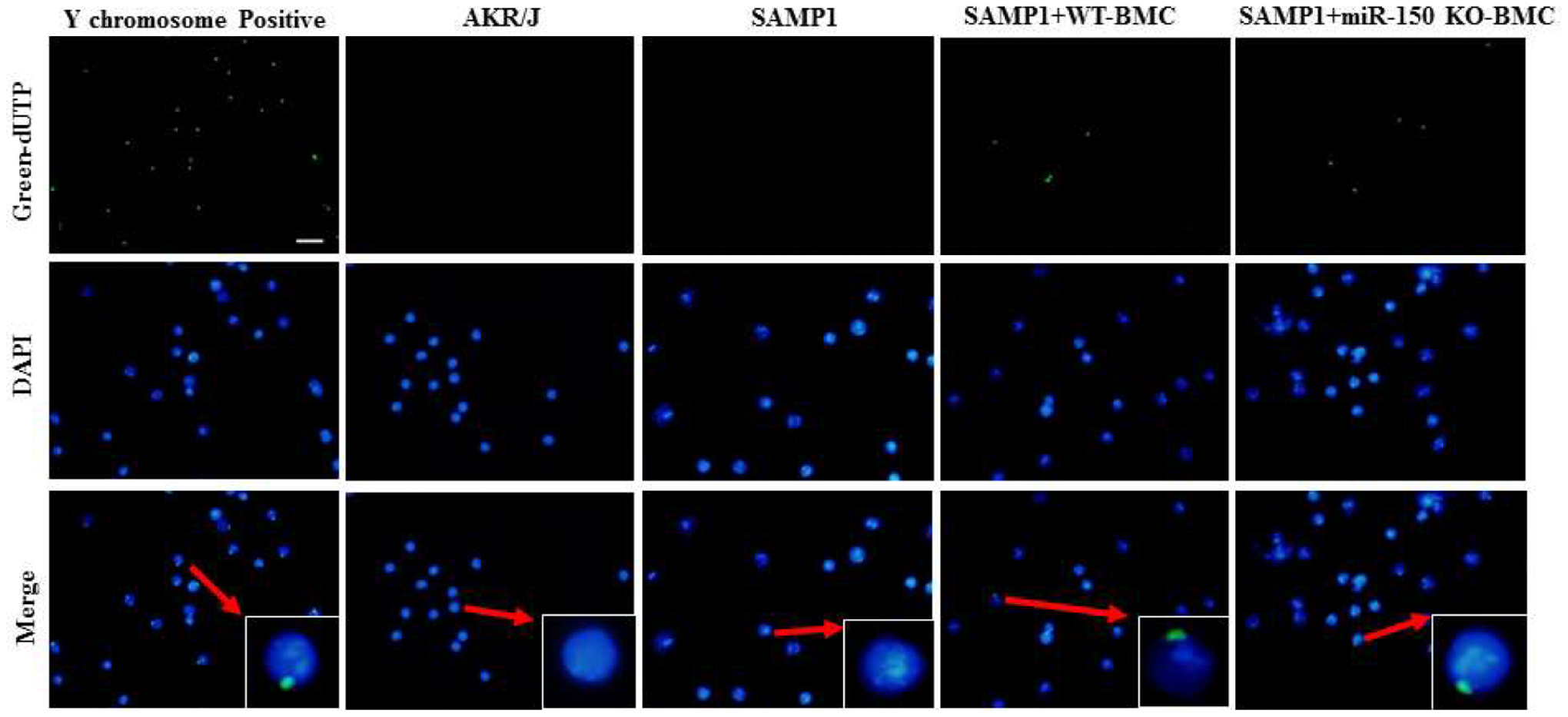
Figure 6.

Representative sections of FISH staining showing that BMCs from male miR-150 KO mice were detected in the BM of female SAMP1 mice at 8 weeks after BMT. The Y chromosomepositive cells (green) and DAPI (blue) for nuclei in the BM were examined at 8 weeks after BMT. Merged images show that Y chromosome is localized in the nucleus. Scale bar indicates 100 μm.
