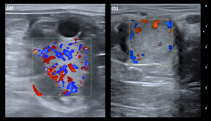
FIGURE 2.

Colour Doppler evaluation of the corpus luteum (CL) in the mare. (a) Colour Doppler ultrasound image of a CL displaying a well‐established homogeneous pattern of vascularization, consistent with an active status of this gland. (b) Colour Doppler ultrasound image of a CL showing an absence of irrigation, indicative of a low functionality
