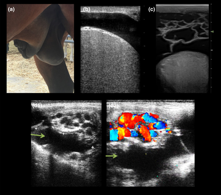
FIGURE 5.

Pathological findings in male reproductive ultrasound. (a) External genitalia from a stallion with heart failure causing ventral oedema. Such tissue swelling could cause problems with the thermoregulation of the testicles as well as mechanical problems when externalizing the penis. (b) B‐mode ultrasound image showing clear anechoic fluid around the testicle, a condition known as hydrocele. (c) B‐mode ultrasound image displaying a cavity with trabeculae between the scrotum and the testicle. This kind image is characteristic of the haematocele. (d) Combination of b‐mode (D1) and colour Doppler ultrasound images of the testicular cord showing a tortuous dilated vein full of fluid (green arrows) but without signs of pulsatility
