
Fig. 4.
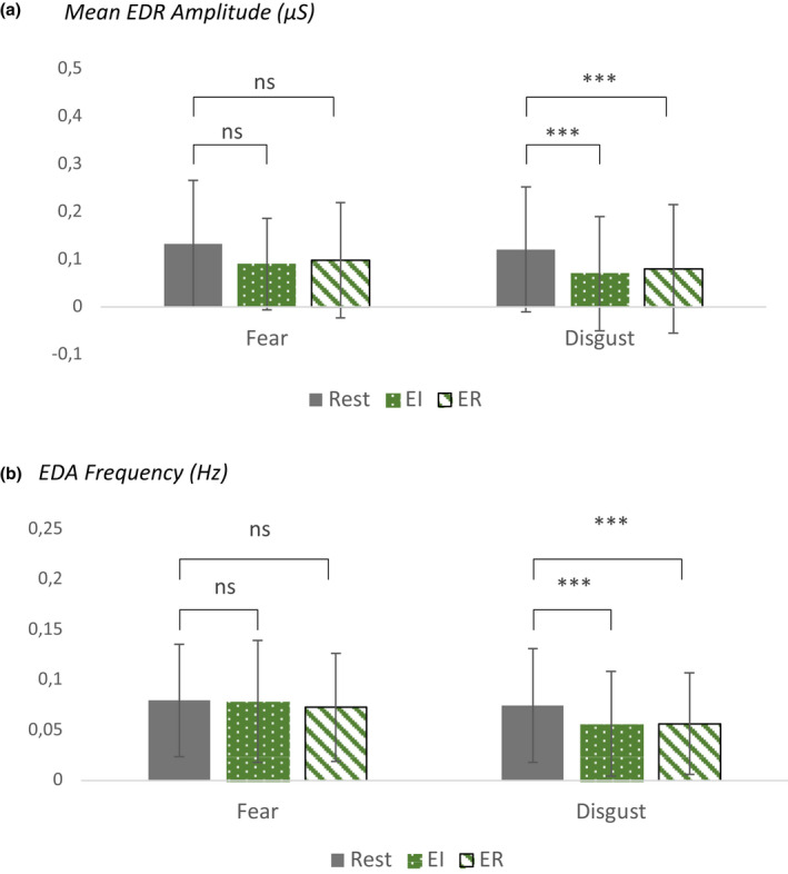
Shows measures of phasic components of electrodermal activity, mean phasic electrodermal response (EDR) amplitude (a) and EDR frequency (b). During EI, we measured increased sympathetic arousal during FEAR, but decreased sympathetic activity during DISGUST. During ER, we observed a reduction of sympathetic activity as reflected in changes in EDA both for FEAR and DISGUST. The two emotions differed significantly only during EI in terms of EDR frequency (shown are mean ± SD). [Colour figure can be viewed at wileyonlinelibrary.com]