Fig. 1.

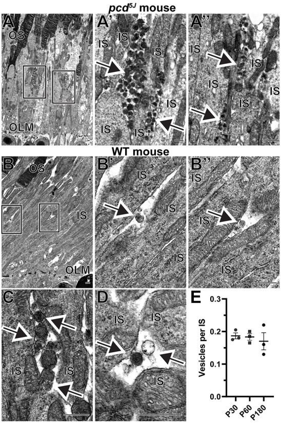
Extracellular vesicles are located next to inner segments of wild-type (WT) and pcd5J mouse photoreceptors. (A) Representative transmission electron microscopy (TEM) images of retinal sections from 22-day-old homozygous pcd5J mice stained with osmium tetroxide. Boxed areas in A are magnified in A′ and A″. (B) A low-magnification TEM image of a WT mouse retinal section stained with osmium tetroxide. Boxed areas are magnified in B′ and B″. (C) An example of a cluster containing several vesicles. (D) An example of two neighboring vesicles with different staining densities. (E) The frequency of vesicles located next to photoreceptor inner segments in WT mice at P30, P60 and P180. The data are expressed as the number of vesicles per inner segment within a region of the 70 nm-thick retinal section containing 200 photoreceptors. Each data point represents an individual mouse; the bars represent mean±s.e.m. One-way ANOVA test revealed no statistically significant differences across ages (P=0.7775). In all panels, arrows point to extracellular vesicles. IS, inner segment; OLM, outer limiting membrane; OS, outer segment. Scale bars: 0.5 µm. A total of four pcd5J and nine WT mice were analyzed.
