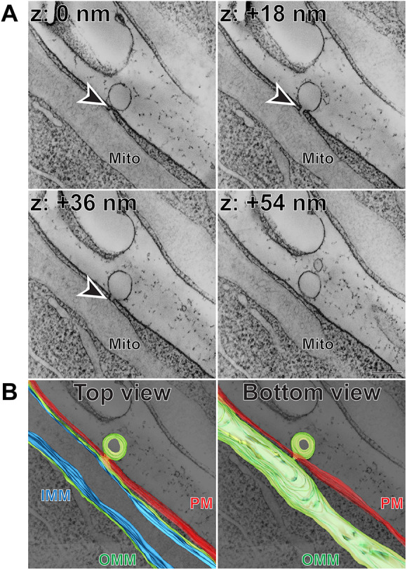
Fig. 3.

Extracellular vesicular structure connected to inner segment mitochondrion through a short membrane tunnel. (A) Representative z-sections at the depths of 0, +18, +36 and +54 nm obtained from a 3D electron tomogram of a 750 nm-thick WT mouse retinal section stained with tannic acid/uranyl acetate. Full tomogram is shown in Movie 3. Arrowheads depict a membrane tunnel connecting the budding vesicle with an inner segment mitochondrion. Tomogram pixel size is 1.5 nm; scale bar: 0.2 µm. (B) The corresponding 3D segmentation in two views: from the top as in A and from the bottom. Full segmentation is shown in Movie 5. IMM, inner mitochondrial membrane (blue); Mito, mitochondrion; OMM, outer mitochondrial membrane (green); PM, plasma membrane (red). The data are selected from a total of 12 tomograms documenting extracellular vesicular structures connected to mitochondrial membranes obtained from three WT mice.
