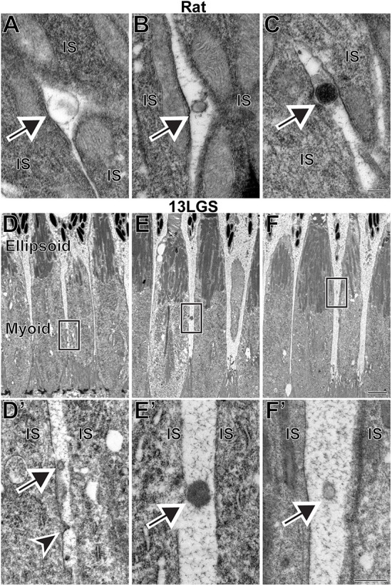
Fig. 5.

Examples of microvesicles located next to the inner segments of WT rat and 13-lined ground squirrel (13LGS) photoreceptors. (A-C) Microvesicles with a variety of staining patterns in TEM images of retinal sections from WT rats stained with tannic acid/uranyl acetate. (D,E,F) Low-magnification TEM images of WT 13LGS retinal sections stained with tannic acid/uranyl acetate. Boxed areas are magnified in D′,E′,F′. In all panels, arrows point to microvesicles; arrowhead points to a microvesicle budding off of a cone inner segment. IS, inner segment. Scale bars: 0.2 µm (A-C); 2 µm (D,E,F); 0.5 µm (D′,E′,F′). A total of four rats and four 13LGS were analyzed.
