FIGURE 6.

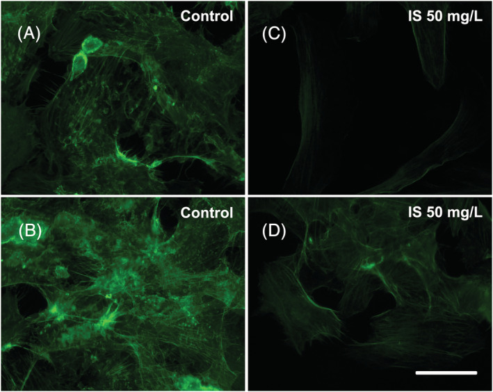
Effects of IS on the actin filament organization: (A,B) On the left, control HMEC‐1 cells observed at 72 h after the start of the treatment. (C,D) On the right, HMEC‐1 cells treated with 50 mg/L IS for 72 h. Images were acquired with ViCo confocal microscope (Nikon). Bar 25 μm
