
Figure 6.
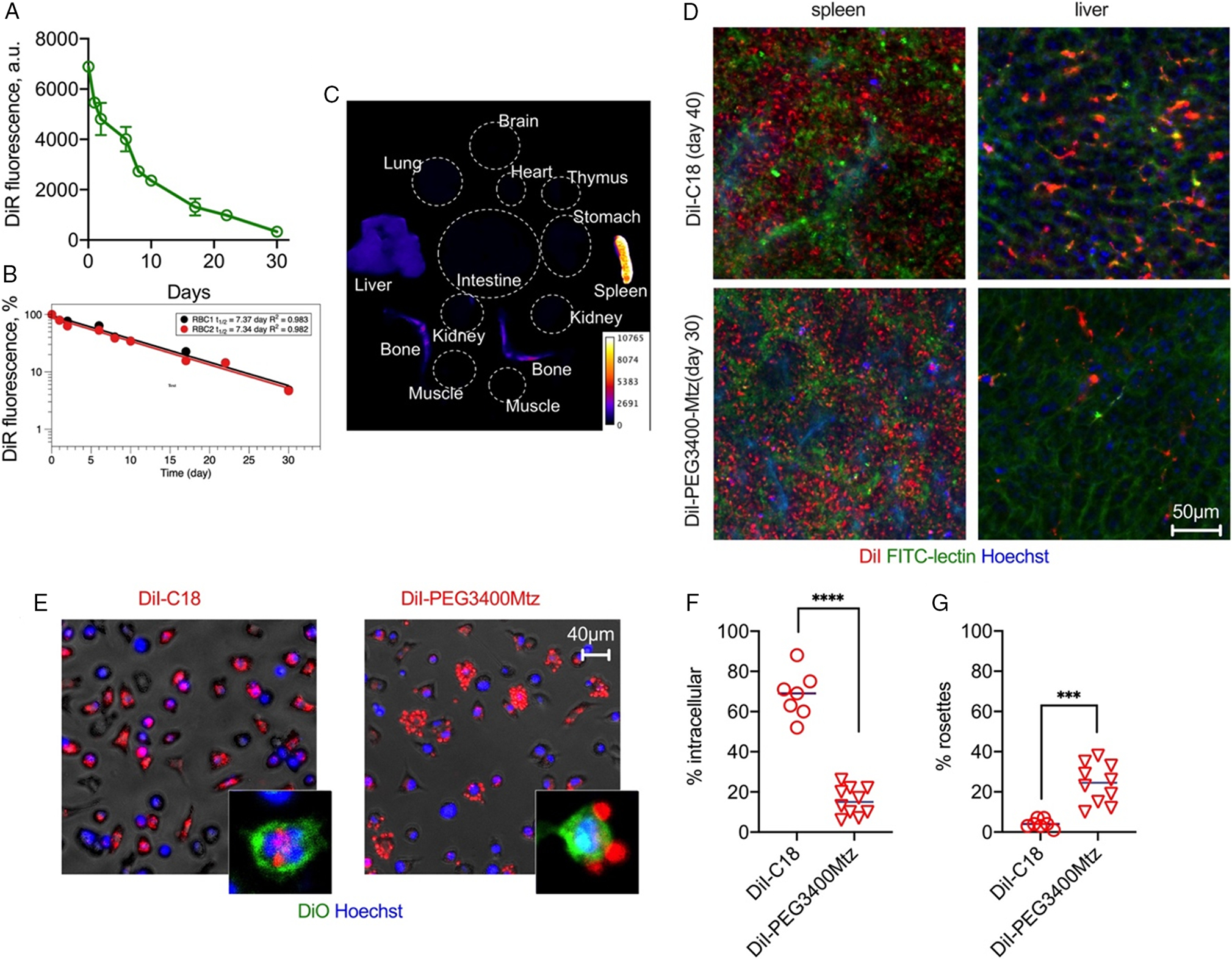
Biodistribution and immune recognition of RBCs. A) RBCs labeled with DiR were injected in BALB/c mice and blood fluorescence was monitored with NIR scanner Li-COR Odyssey. B) One-compartment pharmacokinetic analysis shows long half-life (n = 2 mice). C) Organ biodistribution of DiR fluorescence (pseudocolored) shows predominantly spleen and some liver and bone marrow accumulation. D) Confocal microscopy images of fresh livers and spleens of mice injected with DiI-C18 and DiI-PEG3400Mtz (after in vivo labeling of blood vessels and nuclei with FITC-lectin and Hoechst). The lipid accumulates in extrasinusoidal cells in the spleen and predominantly in sinusoidal cells in the liver (i.e., endothelium and Kupffer cells). DiI-PEG3400Mtz showed low accumulation in the liver. E) RBCs were incubated with fresh peritoneal macrophages for 24 h. DiI-C18 RBCs show mostly intracellular uptake, whereas DiI-PEG3400Mtz RBCs show mostly extracellular rosettes. Insets show confocal images of DiO-labeled macrophages to demonstrate intracellular versus extracellular localization. F) Quantification of percent cells (per field) that contain intracellular DiI. G) Quantification of percent cells (per field) that contain DiI+ rosettes. P-value: ****0.0001; ***<0.001 2-sided t-test, alpha 0.05.