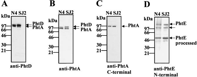
FIG. 3.
Immunoblot analysis of whole-cell lysates of S. pneumoniae. Whole-cell lysates of S. pneumoniae strains N4 and SJ2 were evaluated by immunoblot analysis with mouse antiserum raised against PhtD (residues 21 to 839) (A), PhtA (residues 21 to 816) (B), the C-terminal half of PhtA (residues 386 to 816) (C), and the N-terminal half of PhtE (residues 21 to 484) (D). The reactive bands are indicated by arrows. The molecular masses (in kilodaltons) of the protein standards are indicated.

