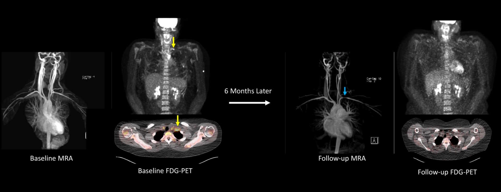
FIGURE 3: Angiographic Progression of Disease in a Patient with Takayasu’s Arteritis.

This 22-year-old female with Takayasu’s arteritis self-discontinued methotrexate/infliximab after approximately 2 years of treatment. Six months later she developed constitutional symptoms, frontal headaches, carotidynia, and left arm claudication. She had significant elevations in acute phase reactants (ESR 104, CRP 85 mg/L). An FDG-PET scan showed severe vascular FDG uptake throughout the aorta and bilateral common carotid arteries with a prominent area of focal inflammation in the left subclavian artery (yellow arrows) on whole-body and axial views. MRA obtained the same date did not show vascular damage. Treatment was re-initiated with excellent clinical response. Six months later, the patient had a repeat MRA showing a new left subclavian artery stenosis (blue arrow) with resolution of vascular PET activity.
