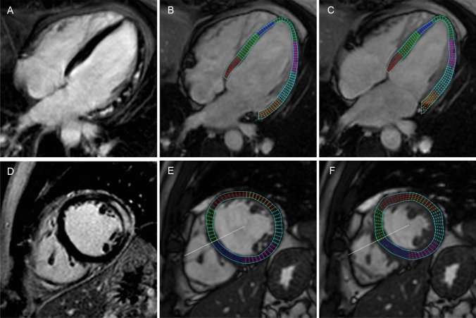
Fig. 2.
Example of Late Gadolinium Enhancement imaging and feature tracking strain analysis by cardiac magnetic resonance in a patient with acute myocarditis. A representative 4 chamber (A) and a short axis midventricular LGE image (D) show extensive involvement of the lateral left ventricular wall. Longitudinal strain analysis is performed in the long axis view as represented by an enddiastolic (B) and endsystolic 4 chamber slice (C). Circumferential and radial strain analysis is assessed using short axis slices as represented by an enddiastolic (E) and endsystolic (F) midventricular slice

