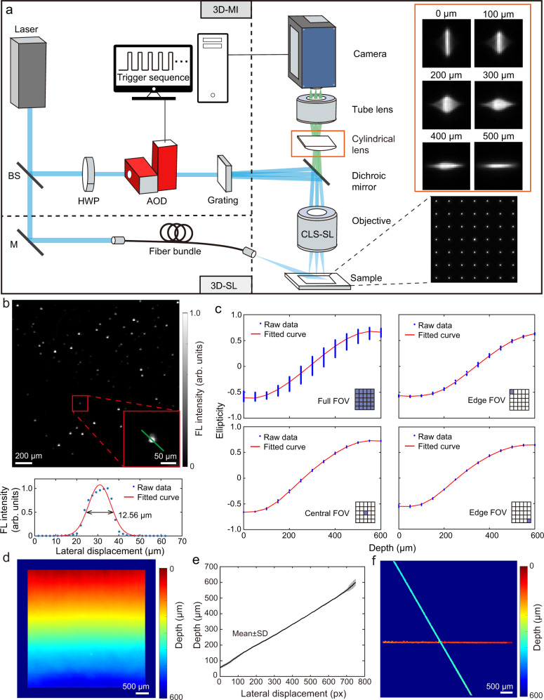
Fig. 1. The integrated volumetric wide-field fluorescence microscopy system based on optical astigmatism and its characterization.
a Schematic of the two spatial localization approaches, which are based on either multifocal illumination (MI) or sparse localization (SL) of individual fluorescence emitters. b Lateral resolution characterization by calculating the full-width-at-half-maximum (FWHM) of the intensity profile of fluorescent beads after Gaussian fitting along the green line. c Representative calibration curves of the PSF ellipticity as a function of depth across the whole FOV (289 illumination spots) and corresponding subregions where 5 illumination spots were selected. d 3D fluorescence image of a tilted microscope slide with linear depth gradient along axial direction. e Averaged line profile along axial direction across the FOV. Data are presented as mean ± SD. f 3D fluorescence image of two crossing tubings separated by approximately 260 μm. BS beamsplitter, M mirror, AOD acousto-optical deflector, HWP half-wave plate, FL fluorescence, FOV field of view, SD standard deviation.

