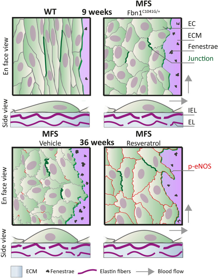
Figure 5.

Overview of endothelial cell dysfunction and elastin sheet disruptions in a mouse model of MFS. Schematic presentation of endothelial cell (EC) organization and extracellular matrix (ECM) structure in the thoracic ascending aorta of WT, MFS, MFS-CTRL, and MFS-RESV mice. In the en face view, differences in EC morphology are shown and two adherens junctions connecting ECs are indicated with a dark green line to highlight the junctional linearity. Moreover, the elastin sheet on which the ECs are situated is drawn with occasional fenestrae. Expression of p-eNOS on the EC membrane is shown as red lines at the age of 36 weeks. In the side view, EC morphology is shown, and the fenestrae are now indicated as breaks in the internal elastic lamina (IEL). Part of the aortic media is shown with its ECM and a medial elastic lamina (EL) (vSMCs are not depicted). Arrows indicate blood flow direction.
