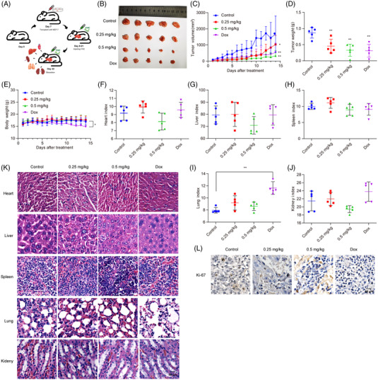
FIGURE 1.

The in vivo anti‐tumour effect of YHC on human breast cancer MCF‐7 cells by using xenograft models. (A) The experimental flow chart for evaluating the anti‐tumour effect of YHC. (B) The tumour issues were excised and photographed after treatment with YHC for 14 days. (C) Tumour volumes were measured using calipres for each group every day. (D) Tumour weights were detected in each group. (E) The body weights of animals were recorded for each group during 14 days. (F–J) The organ index of nude mice was calculated for heart (F), liver (G), spleen (H), lung (I) and kidney (I), respectively. (K) Representative histomorphological changes of the organs were assessed after H&E staining. (L) YHC could decrease Ki‐67 expression in tumour tissues by immunohistochemistry assay. Statistical analysis was performed on the data as the mean ± SD (n = 5). * p < .05, ** p < .01 versus control group.
