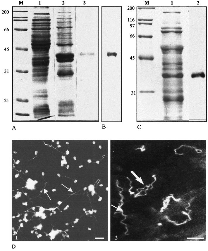
FIG. 1.
(A) SDS-PAGE analysis of E. coli-expressed recombinant EcPTP2 (Coomassie blue staining). Lanes 1 and 2, total E. coli lysate before (lane 1) and after (lane 2) IPTG induction; lane 3, recombinant EcPTP2 after purification on an Ni-NTA column; lane M, molecular mass standards in kilodaltons. (B) Immunoblotting reactivity of MAb Ec102 with E. coli proteins 4 h after IPTG induction. The blot was probed with a 1:10,000 dilution of MAb Ec102 and developed using ECL (Amersham). (C) Lane 2, immunoblot of E. cuniculi whole-cell homogenates probed with anti-recombinant EcPTP2 antiserum. Lane 1, total E. cuniculi proteins stained with Coomassie blue. (D) Indirect immunofluorescence of E. cuniculi spores with extruded polar tubes (arrows). (1) Labeling with an antiserum against total proteins; (2) specific labeling of polar tubes with anti-recombinant PTP2. Bars, 5 μm.

