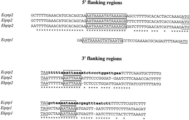
FIG. 3.
Alignments of ptp1 and ptp2 gene 5′ and 3′ flanking regions showing conserved signals. Start and stop codons are underlined. Identical nucleotides are indicated by asterisks. The AT-rich consensus sequence in the 5′ region and potential site of polyadenylation are boxed. Partial E. cuniculi cDNAs with short 3′ UTRs (less than 30 nucleotides) are in boldface lowercase letters.

