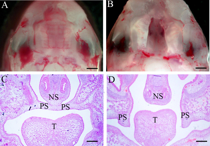
Figure 1.

Morphology and H&E of palate shelf tissues at E16.5. (A, C) The palatal shelf contacted the midline and fused through the formation of the midline epithelial seam (MES) in the mid-anterior region of a control embryo. (B, D) Unfused, separated palatal shelf from an embryo of RA-treated mouse. (A, B) Morphological specimens (magnification ×100, scale bar 100 μm); (C, D), H&E staining results (magnification ×40, scale bar 200 μm). PS, palatal shelf; T, tongue; NS, nasal septum; H&E, hematoxylin, and eosin.
