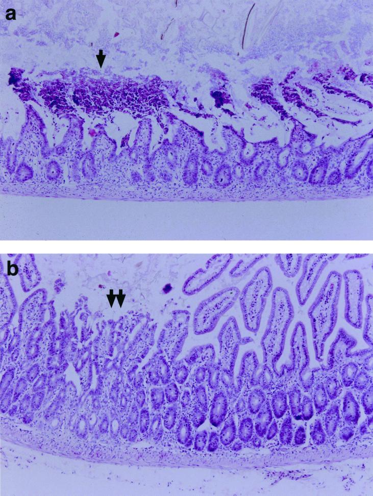
FIG. 2.
Representative hematoxylin and eosin (H&E)-stained sections from the small intestines of (a) WT and (b) IL-5−/− male mice. Mice were infected with 10 T. gondii cysts by the oral route, and their intestines were removed for examination at 8 days postinfection. There was extensive necrosis of the small intestinal villi of WT mice (a, arrow). In contrast the small intestine of the IL-5−/− mice showed mild blunting of the villus architecture and focal necrosis (b, double arrow).

