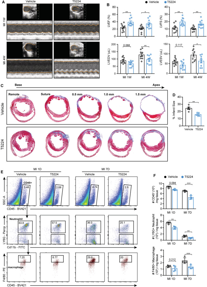
Figure 8. The selective Fos/AP‐1 inhibition mitigated cardiac leukocyte infiltration and cardiac dysfunction.

A, Wild‐type mice were subjected to MI surgery and randomly treated with Fos/AP‐1 inhibitor (T5224) or its vehicle for 7 consecutive days, then the echocardiography was used to compare the cardiac function at 1 and 4 weeks post MI. B, Quantification of A (n=9 for each group; 2‐way ANOVA followed by least significant difference test). C, Masson trichrome staining of vehicle‐ or T5224‐treated mice to determine the infarct size at 4 weeks post MI. D, Quantification results of C (n=5 for each group; Student's t‐tests). E, Number of leukocytes, neutrophils, and macrophages at 1 and 7 days after MI in vehicle‐ or T5224‐treated mice, as measured by flow cytometry analysis. F, Quantification results of panel E (n=4 for each group; 2‐way ANOVA followed by least significant difference test). AP‐1 indicates activator protein 1; LVEDV, left ventricular end diastolic volume; LVEF, left ventricular ejection fraction; LVESV, left ventricular end systolic volume; LVFS, left ventricular fraction shortening; and MI, myocardial infarction.
