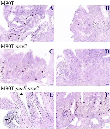
FIG. 6.
Immunoperoxidase labeling of serotype 5 somatic antigen by anti-LPS monoclonal immunoglobulin G. (A, C, and E) Sections of PPs (A and C) and villi (E) from animals infected i.g. at day 1 p.i; (B, D, and F) sections of PPs of animals at day 7 p.i. (A and B) Sections from guinea pigs infected with M90T; (C and D) sections from guinea pigs infected with M90T aroC; (E and F) sections from guinea pigs infected with M90T purE aroC. Bars, 50 μm (A, B, C, D, and F) and 20 μm (E). The inset in panel E shows the tip of a villus in which LPS material can be observed. Arrowheads point to bacteria or bacterial components present within villi.

