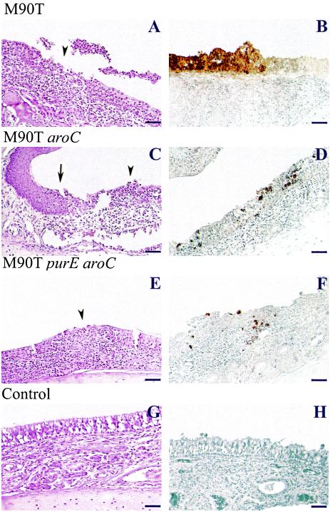
FIG. 7.
Infection of guinea pig nasal mucosa with M90T (A and B), M90T aroC (C and D) and M90T purE aroC (E and F) at day 1 p.i; (G and H) nasal mucosa from an uninfected animal. (A, C, E, and G) Hematoxylin-eosin stained tissue sections; (B, D, F, and H) immunoperoxidase labeling of serotype 5 somatic antigen by anti-LPS monoclonal IgG. Arrowheads (A, C, and E) point to areas of necrosis of the associated simple epithelium. Arrow (C) indicates the transition between the stratified squamous epithelium and the simple epithelium where areas of necrosis are observable. Bar, 20 μm.

