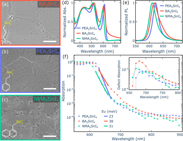
Figure 1.
Scanning electron microscope (SEM) images show the crystallization morphologies of three different perovskites: BA2SnI4 (a), PEA2SnI4 (b), and NMA2SnI4 (c) (scale bar = 2 μm). The absorption (d) and photoluminescence spectra (e) for these three perovskites are also presented. (f) Photothermal deflection spectra comparing the Urbach tails of these three materials to the corresponding Urbach Energy (EU). The inset highlights the defect absorption by subtracting the fitted Urbach tail from the pristine spectra. All results correspond to room temperature measurements.

