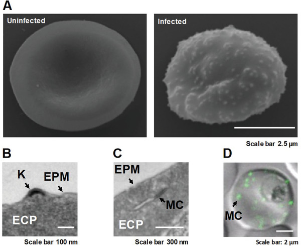
Figure 2.

Reorganization of the host RBC by the malaria parasite Plasmodium falciparum. A) Scanning Electron Microscopy (SEM) images of an uninfected and an infected RBC show the marked changes exerted by the parasite. The RBC loses its biconcave shape while the parasite grows. Small protrusions, so‐called knobs, are presented on the surface of the infected RBC. The knobs serve as presentation platforms for the cytoadherent parasite protein PfEMP1. B) Transmission Electron Microscopy (TEM) image of a thin sectioned infected RBC shows the electron density of the knob. The successful establishment of knobs depends on the Maurer's clefts, a novel secretory organelle generated by the parasite in order to facilitate protein trafficking in the usually cell‐organelle‐deprived RBC C). The Maurer's clefts consist of several single cisternae or a few stacks of several cisternae which are usually spread throughout the infected RBC. The morphology of the organelle depends on the parasite strain. D). The confocal image shows the location of the Maurer's clefts via the GFP‐tagged PfSBP1 (P. falciparum skeleton binding protein 1) which attaches the cisternae of the newly generated Maurer's clefts to actin filaments that have been mined from the membrane skeleton of the RBC by the parasite. Reproduced under the terms of the Creative Commons CC‐BY license.[ 12 , 325 ] Copyright 2019, The Authors. Published by Wiley‐VCH.
