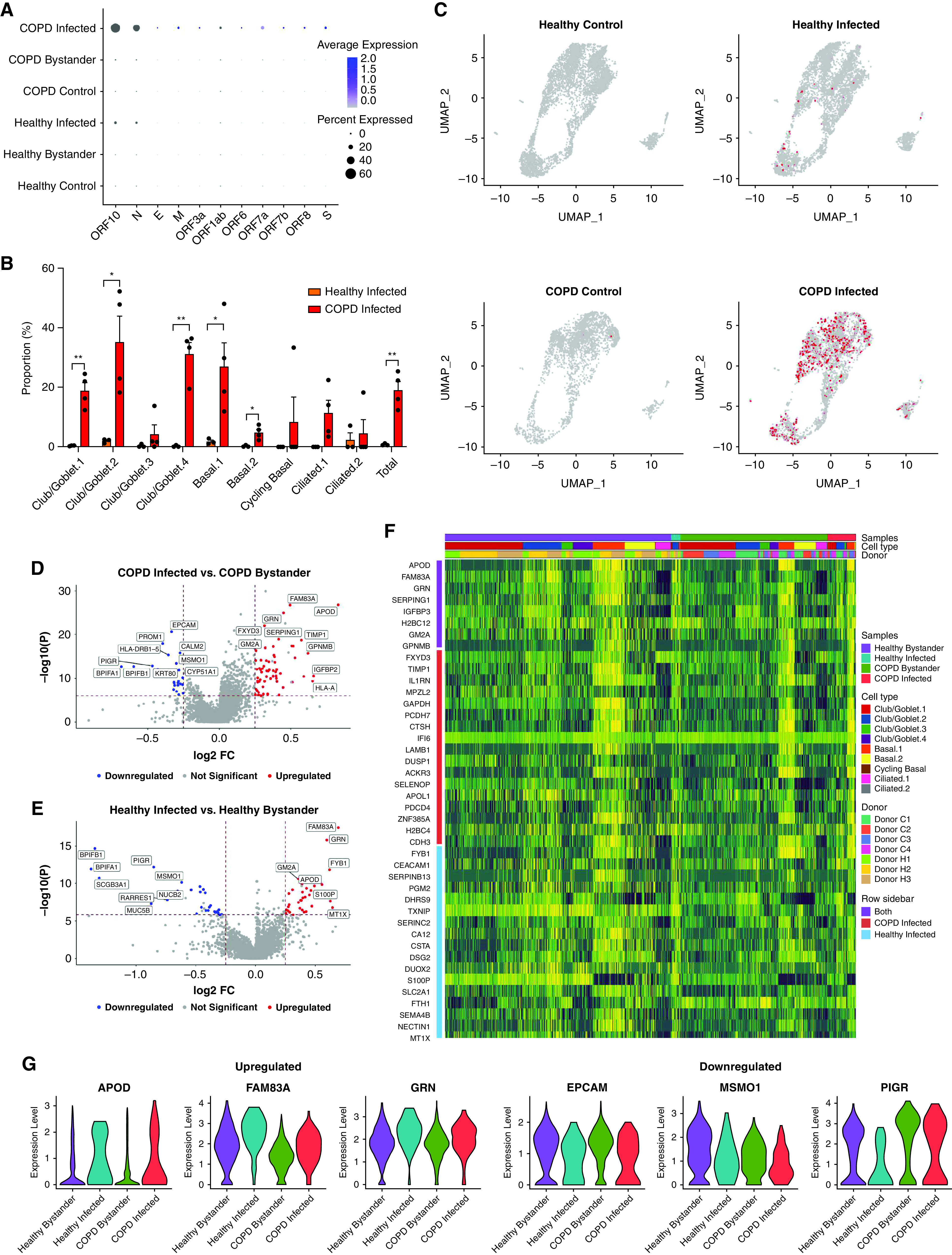
Figure 2.

Differential gene expression analysis between infected and bystander cells in infected chronic obstructive pulmonary disease (COPD) and healthy primary bronchial epithelial cells. (A) Signature plots of severe acute respiratory syndrome coronavirus 2 (SARS-CoV-2) markers. (B) Proportion of infected healthy (orange) and COPD (red) cells in each cell cluster. (C) Expression of SARS-CoV-2 markers in each of the different groups. (D) Volcano plot comparison of infected and bystander COPD cells. (E) Volcano plot comparison of infected and bystander healthy cells. (F) Heatmap of the top 25 genes in infected compared with bystander healthy and COPD cells. (G) Violin plots of three common upregulated and downregulated genes in infected healthy and COPD cells compared with their respective bystander cells. Statistical differences between the groups are indicated whereby *P ⩽ 0.05 and **P ⩽ 0.01. FC = fold change; UMAP = uniform manifold approximation and projection.
