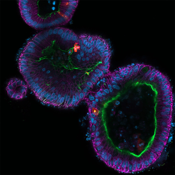
Figure 2.

Fluorescent micrograph of multiple human intestinal organoids infected with rotavirus. The organoids were fixed and stained for E-cadherin (magenta), villin (green), nuclei (blue), and rotavirus (red). The image was acquired on a Zeiss LSM980 confocal microscope.
