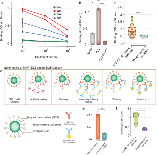
Figure 4.

Immunological evaluation and serological application of epitope‐scaffolds. a) Sera from mice immunized with epitope‐scaffolds were tested by ELISA for binding to each epitope‐scaffold. Data include two animals per group. b) Comparison of ED2 immunized mice serum response to ED2, ED2 control, and spike protein. c) Anti‐epitope‐1 IgG antibodies were detected in both negative controls (pre‐pandemic samples) and COVID‐19 patient samples using ED2. Data represent violin plots and individual data points. n = 5 samples for pre‐pandemic samples and n = 25 samples for COVID‐19 patient samples. d) Schematic representation of ELISA based on ED2‐decorated magnetic nanoparticle (MNPs) for the detection of epitope‐1 specific IgG antibodies. e) Anti‐epitope‐1 IgG antibodies were tested in negative controls and COVID‐19 patient samples using ED2 and by employing MNP‐based ELISA. f) Anti‐epitope‐1 IgG antibodies were tested in negative controls and COVID‐19 patient samples using ED2 and by employing MNP‐based ELISA. Data represent violin plots and individual data points. n = 4 samples for pre‐pandemic samples and n = 10 samples for COVID‐19 patient samples.
