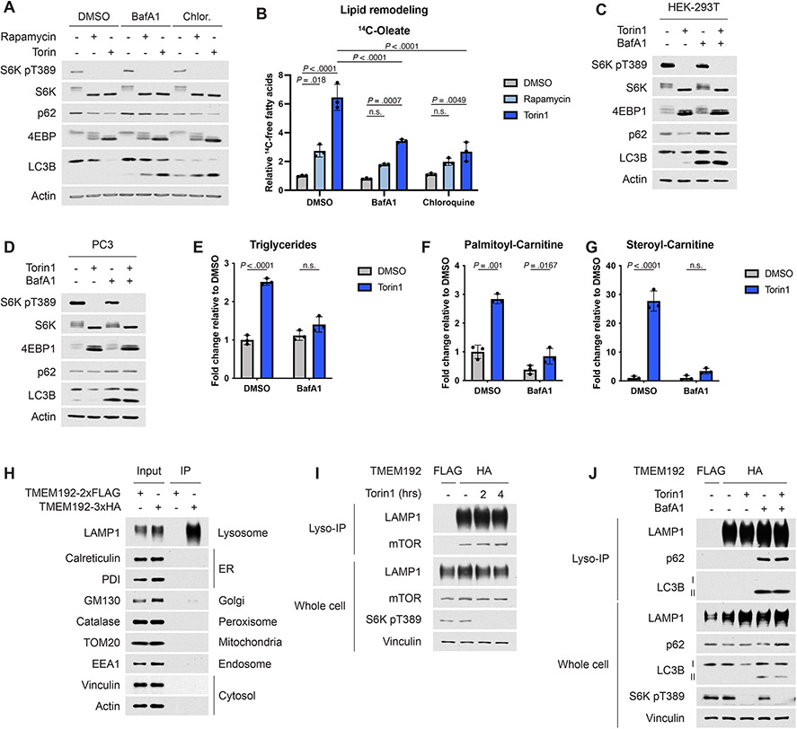
Extended Data Fig. 5.
(A,B) Immunoblot (A) and fatty acids (B) corresponding from the experiment shown in Figure 5A. Fatty acids quantification is graphed as mean ± SD relative to vehicle-treated cells, n=3.
(C,D) Immunoblots corresponding to the experiments shown in Figures 5B,C.
(E-G) Triglyceride and acyl-carnitine levels from the experiment shown in Figures 5D-G.
(H) Immunoblot for organelle markers in whole cell lysates or in HA-tag immunopurified lysosomes (IP) from HEK-293T cells expressing TMEM192-2xFLAG or 3xHA.
(I) Immunoblot corresponding to the experiment shown in Figures 5K,L.
(J) Immunoblot corresponding to the experiment shown in Figures 5M,N. LC3B is indicated in its unmodified (LC3B-I) and lipidated (LC3B-II) forms.
Statistical analysis by two-way ANOVA (B,E-G). n.s., not significant.

