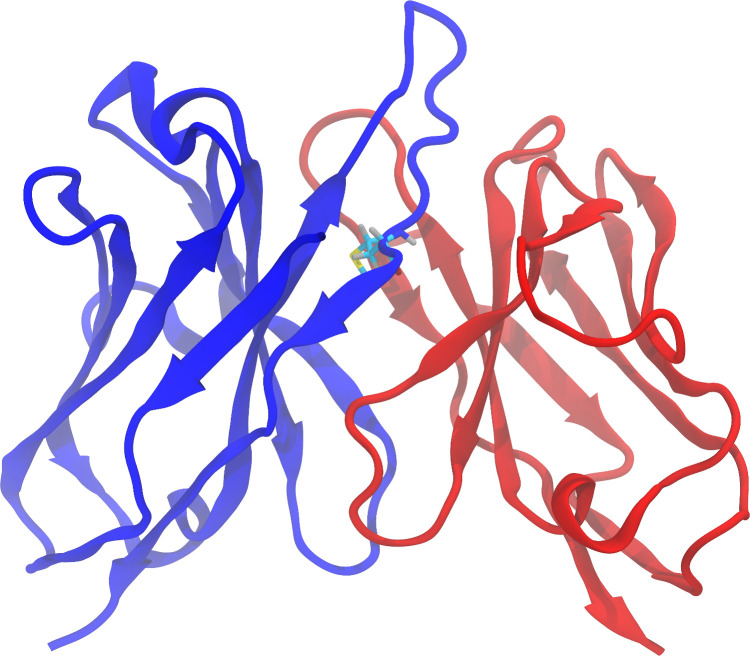
Fig 1. Three-dimensional structure (homology model) of the Fv region of the antibody Lirilumab.
The heavy chain and the light chain are represented in blue and red, respectively. Methionine 100F, located at the end of the H3 loop, is shown with carbon atoms colored in cyan, oxygen in red, hydrogen in gray, nitrogen in blue and sulfur in yellow.

