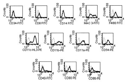
FIG. 3.
Flow cytometric analysis of lung dendritic cells cultured for 7 days. When CD11c+ lung cells were cultured for 7 days in cRPMI containing recombinant murine GM-CSF, all cells remained CD11c+ and were similar to the R1 population in Fig. 2. Levels of cell surface expression for different markers on 7-day culture of lung dendritic cells were analyzed. Lung dendritic cells were incubated with MAbs against CD34, CD8, CD14, MAC3, F4/80, CD11c (N418), CD11c (HL3), CD11b CD11a CD54, CD40, CD80, and CD86 antigens. The histogram for each specific MAb was overlaid against its corresponding isotype control (dashed line). Data are shown for differentiation markers (top row), integrins and adhesion molecules (middle row), and costimulatory molecules (bottom row). PE, phycoerythrin.

